Blog Author: Archita KhaireBreast cancer is the most common cancer worldwide. Last year 2.3 million women were diagnosed with breast cancer, and 685,000 deaths were reported. The National Cancer Institute states that more than $500 million is spent yearly on breast cancer research. Even with the significant investment, approximately 43,000 women in the U.S. are expected to die in 2021 from breast cancer.
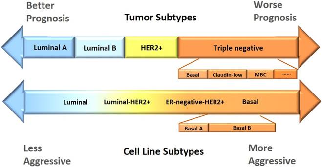
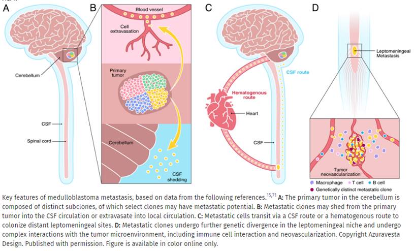
Based on the human epidermal growth factor receptor 2 (Her2), progesterone receptor (PR), and estrogen receptor (ER), breast cancer is categorized into four molecular subtypes: Blog Author: Archita KhaireMedulloblastoma is the most prevalent brain tumor type in children. It is also called a cerebellar primitive neuroectodermal tumor (PNET)—that starts in the region of the brain at the base of the skull, called the posterior fossa. In the United States, about 400 children are diagnosed with #medulloblastoma every year. The survival rate of Medulloblastoma is about 70%.
Blog Author: Archita KhaireWhat is a brain tumor?
A brain tumor occurs as a result of an abnormal growth or spread of cells from within the brain or its supporting tissues that can damage the brain. Glioblastoma (also known as GBM) is the most aggressive type of brain tumor. It accounts for 48 percent of all primary malignant brain tumors. As per National Brain Tumor Society more than 10,000 individuals in the United States lose their life because of #glioblastoma every year. Prognosis of the patients with #GBM is very low (< 15 months). Let's look at different stages of GBM where A.I. is playing a critical role in helping healthcare professionals. Diagnosis Brain tumor diagnosis is typically a two-step process: MRI & Biopsy Magnetic resonance imaging (MRI) Doctors use MRI to detect the presence of tumors in the brain. #MRI is non-ionizing, non-invasive technique and provides good spatial and temporal resolution. But the segmentation and delineation of MRI images is time consuming and accuracy depends on expertise of the person analyzing the images. Deep learning algorithms are now helping radiologists to accurately identify and segment the tumors in MRI scans. Multiple images of brain are acquired using volumetric MRI. T1-weighted and T2-weighted are the most common MRI sequences. Fluid Attenuated Inversion Recovery (Flair) is also a frequently used sequence in brain tumors. Different types of MRI sequences help with delineation of the GBM. Edema, a fluid like collection surrounding the tumor cells is best visible in the Flair and T2 sequence. Necrotic region (dead cells) and enhancing tumor is very well seen in T1 post contrast sequence. |
Page HitsAuthorArchita Archives
January 2023
Categories |


 RSS Feed