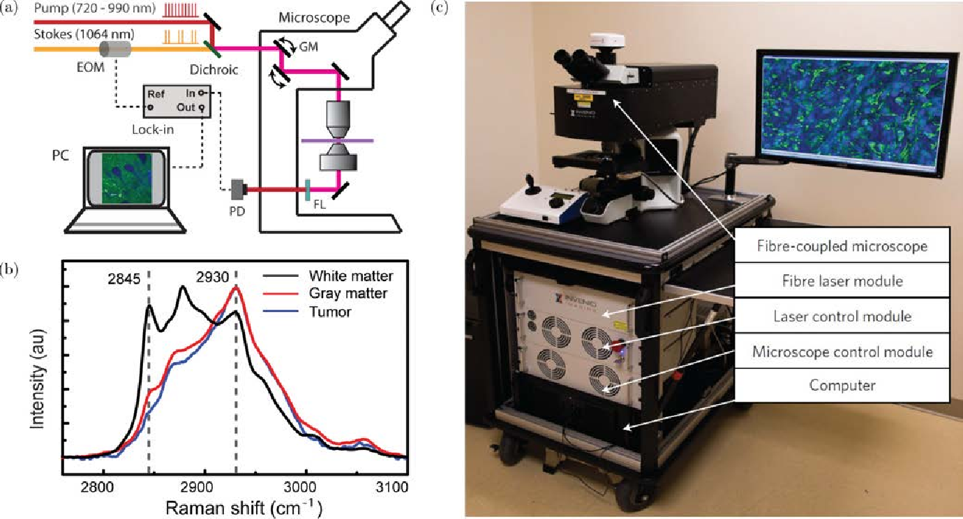
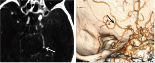
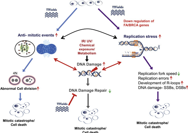
Blog Author: Archita KhaireWhat is a brain tumor? A brain tumor occurs as a result of an abnormal growth or spread of cells from within the brain or its supporting tissues that can damage the brain. Glioblastoma (also known as GBM) is the most aggressive type of brain tumor. It accounts for 48 percent of all primary malignant brain tumors. As per National Brain Tumor Society more than 10,000 individuals in the United States lose their life because of #glioblastoma every year. Prognosis of the patients with #GBM is very low (< 15 months). Let's look at different stages of GBM where A.I. is playing a critical role in helping healthcare professionals. Diagnosis Brain tumor diagnosis is typically a two-step process: MRI & Biopsy Magnetic resonance imaging (MRI) Doctors use MRI to detect the presence of tumors in the brain. #MRI is non-ionizing, non-invasive technique and provides good spatial and temporal resolution. But the segmentation and delineation of MRI images is time consuming and accuracy depends on expertise of the person analyzing the images. Deep learning algorithms are now helping radiologists to accurately identify and segment the tumors in MRI scans. Multiple images of brain are acquired using volumetric MRI. T1-weighted and T2-weighted are the most common MRI sequences. Fluid Attenuated Inversion Recovery (Flair) is also a frequently used sequence in brain tumors. Different types of MRI sequences help with delineation of the GBM. Edema, a fluid like collection surrounding the tumor cells is best visible in the Flair and T2 sequence. Necrotic region (dead cells) and enhancing tumor is very well seen in T1 post contrast sequence. The convolutional neural network, or CNN is the most commonly used machine learning algorithm for image classification. #CNN algorithm segments the MRI images pixel by pixel by applying filters to extract the features. These features are used to label each pixel on MRI as either no tumor, tumor core, edema. This quantitative analysis of medical images to characterize the images is known as Radiomics It is enabling healthcare professionals to conduct clinical trials with greater accuracy and speed. Biopsy If MRI indicates presence of dead tissue in the brain, then sample tissue taken out using biopsy and analyzed by a pathologist to determine the type of brain cancer. Tissue #biopsy is an invasive and time intensive process involving small surgery for tissue extraction. Now researchers have shown that combination of advanced imaging techniques called Stimulated Raman Histology (SRH) and A.I. can be used to quickly analyze images of brain tumor biopsies directly in the operating room. Efforts are being made to develop advanced machine learning algorithms to detect traces of cancer in liquid biopsy. Image Credit: Yang, Yifan et al. “Stimulated Raman scattering microscopy for rapid brain tumor histology.” Journal of Innovative Optical Health Sciences 10 (2017): 1730010. Sometimes doctors recommend additional tests for enhanced diagnosis of the tumor. Computed Tomography (CT) scan is used to find bleeding and enlargement of the fluid-filled spaces in the brain. #ctscan exposes patients to harmful x-rays. AI is now enabling radiologists to conduct low-dose CT imaging to alleviate concerns around radiation exposure. Algorithms have potential to artificially improve the quality of images thus allowing patients to spend lesser time in the imaging room. For example Generative Adversarial Network (#GAN) takes under-sampled image and reconstructs enhanced images using Simultaneous Algebraic Reconstruction Technique. TrueFidelity is one such CT image reconstruction product developed by GE company. Positron emission tomography (PET) scan detects actively dividing cancer cells that show higher levels of absorption of small radioactive substances (fluorodeoxyglucose-18) injected in the patient's body. Although PET offers better sensitivity than other types of scans, its image resolution and signal to noise ratio (SNR) are still low due to various physical degradation factors and low number of coincidences. Deep learning algorithms are being developed to improve PET signal detection, data de-noising, data corrections , image reconstruction, image processing and quantification. Cerebral arteriogram, is type of an x-ray taken after a special dye called a contrast medium is injected into the main arteries of the patient’s head. This test helps doctors detect life-threatening cerebral aneurysms, weak spots in blood vessels in the brain which can balloon out and fill with blood. The test is known as angiogram. CNN algorithms are being developed to analyze the CT angiography images for detecting cerebral aneurysms. Left: CT angiogram showing an aneurysm of 2 mm maximum diameter on the left posterior cerebral artery (arrow). Right: volume-rendered 3D reconstruction image. The aneurysm was missed in the initial report but successfully detected with the deep-learning algorithm. (Image Credit: Radiological Society of North America) Spinal tap is used to check cerebrospinal fluid (CSF) for presence of tumor cells. Machine learning code is now available to detect protein signatures in #CSF to identify presence of tumor cells in very early stages of cancer. Myelogram is recommended to find out if the tumor has spread to the spinal fluid. You can find more information here Molecular testing of the tumor to identify specific genes, proteins unique to the tumor. Over the years many AI techniques have been developed in Molecular diagnosis. Electroencephalography (EEG) is used to monitor for possible seizures. IBM has developed AI algorithm that can detect seizures with 98.4% accuracy using #EEG. Recently a wearable device was developed with a machine learning model to detect seizures in advance. Treatment With so much data being collected during diagnosis phase, it is becoming possible to build personalized cancer treatment for GBM. Surgery continues to be a mainstream treatment for GBM. A craniotomy is the most common type of operation where the neurosurgeon cuts out an area of bone from your skull to remove the tumor. Neuroendoscopy is another type of surgery in which the surgeon puts the endoscope through the hole in the skull to remove the tumor. So far AI in brain surgery has mostly remained in research papers. As soon as skull is cut open, brain starts moving to equalize intracranial and atmospheric pressure thus requiring real time adjustments to locate and remove the tumor. Removing any unwanted brain tissue could result in serious neurologic deficits. Because of these challenges autonomous robotic surgery on brain is still not a reality. AI augmented robots are being built which could assist neurosurgeons during live surgery. Franka Emika and ZEISS KINEVO 900 are some of the advance surgeon controlled robots that are vastly improving the precision of #neurosurgery. Radiation therapy treats the GBM patients by destroying tumor cells with high-energy x-rays. #External-beam is most common type of radiation therapy. Conventional radiation therapy, 3-dimensional conformal radiation therapy (3D-CRT), Intensity modulated radiation therapy (IMRT),Proton therapy and Stereotactic radio-surgery are the different ways External-beam therapy is directed. Lot of research is being done in radiation oncology to integrate AI with radiation therapy to achieve a personalized and precise approach to cancer treatment using radiation, known as precision oncology. Chemotherapy involves treating GBM patient with drugs to destroy tumor cells. there are possible side effects of #chemotherapy that include fatigue, risk of infection, nausea and vomiting, hair loss, loss of appetite and diarrhea. AI is now used to predict the tolerance of chemotherapy drugs, so as to personalize the chemotherapy dose for patient. Alternating electric field therapy, also known as Tumor Treating Fields (TTFields or TTF) disrupts tumor cell mitosis thus causing cancer cell death. Image Credit: UTSouthWestern Medical Center
Unlike chemotherapy and radiation, #TTFields therapy does not cause side effects like pain, nausea, fatigue. Machine learning algorithms are being used for targeted delivery of #TTF for effective treatment of GBM. Future Scope Glioblastoma is one of the most difficult cancer to treat. Early diagnosis of GBM improves the survival rate. Artificial intelligence is making MRI and biopsy more powerful thus helping doctors decide course of treatment. Next challenge is to find the personalized drugs and non-invasive surgeries that could help patients with quick recovery. References
Comments are closed.
|
Page HitsAuthorArchita Archives
January 2023
Categories |


 RSS Feed